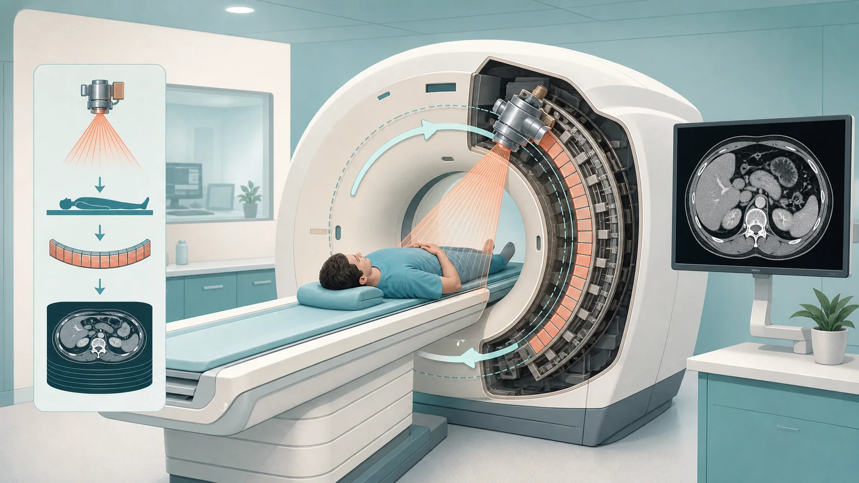
Free ArticleArticle
Medical Imaging Foundations
What medical imaging is for, why exams are ordered, and how an image becomes part of the patient record.
Read Article →A focused free-learning hub where you can watch concise video lessons, download practical reference PDFs, and use the blog as a deeper study resource alongside the main site.
Move between short video lessons, downloadable reference material, foundation articles, and blog content without leaving the same learning environment.
Start with free articles that build the clinical, technical, and workflow language new learners need first.
Helpful open resources, kept organized and easy to expand as the academy grows.
Practical PDFs, diagrams, and workflow guides in one place to build a reusable personal reference library.
Four short articles that introduce the language of medical imaging - what imaging is for, the clinical terms you'll hear every day, how systems fit together, and how change is managed in a live clinical environment. They're high-level on purpose: orientation before the deeper courses.
What medical imaging is for, why exams are ordered, and how an image becomes part of the patient record.
Read Article →
The clinical language behind exam names and image display: anatomy, physiology, directional terms, body planes, and X-ray projections.
Read Article →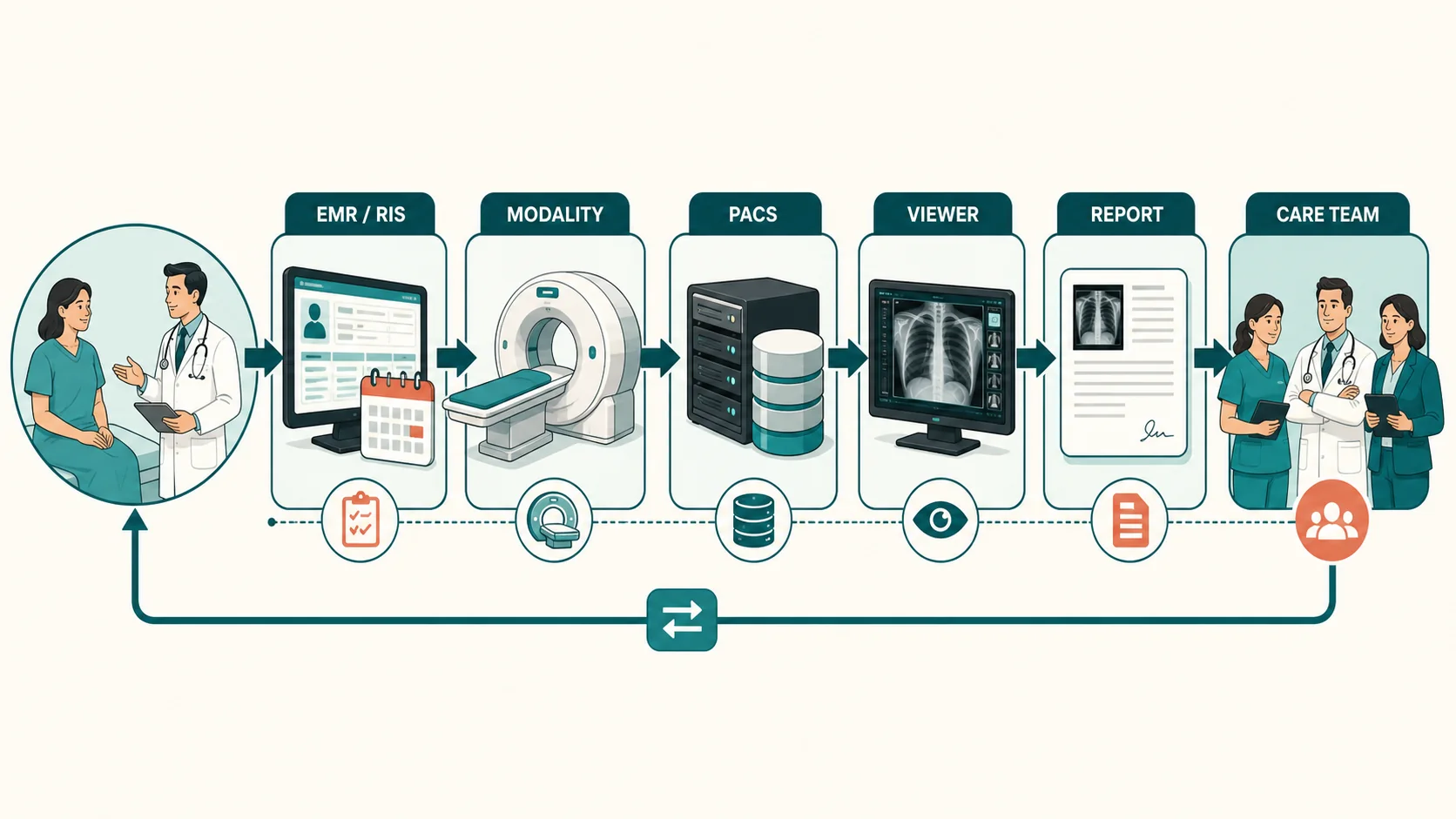
How images, data, standards, systems, and people connect - PACS, viewers, DICOM, HL7, worklists, and reporting at a glance.
Read Article →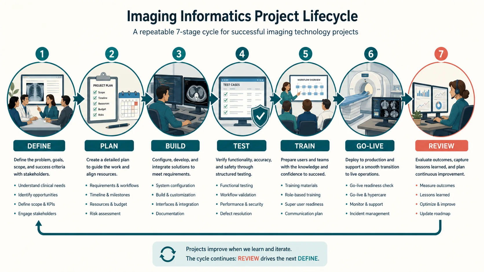
How upgrades, workflow changes, testing, go-live, and support are planned and controlled in imaging projects.
Read Article →The videos give you quick momentum; the blog adds broader explanation, practical examples, and thought pieces that connect technical concepts to real imaging operations.

A workflow-focused PDF to use as a practical companion while learning how imaging orders, modalities, DICOM, PACS, and reporting fit together across the real operational path.
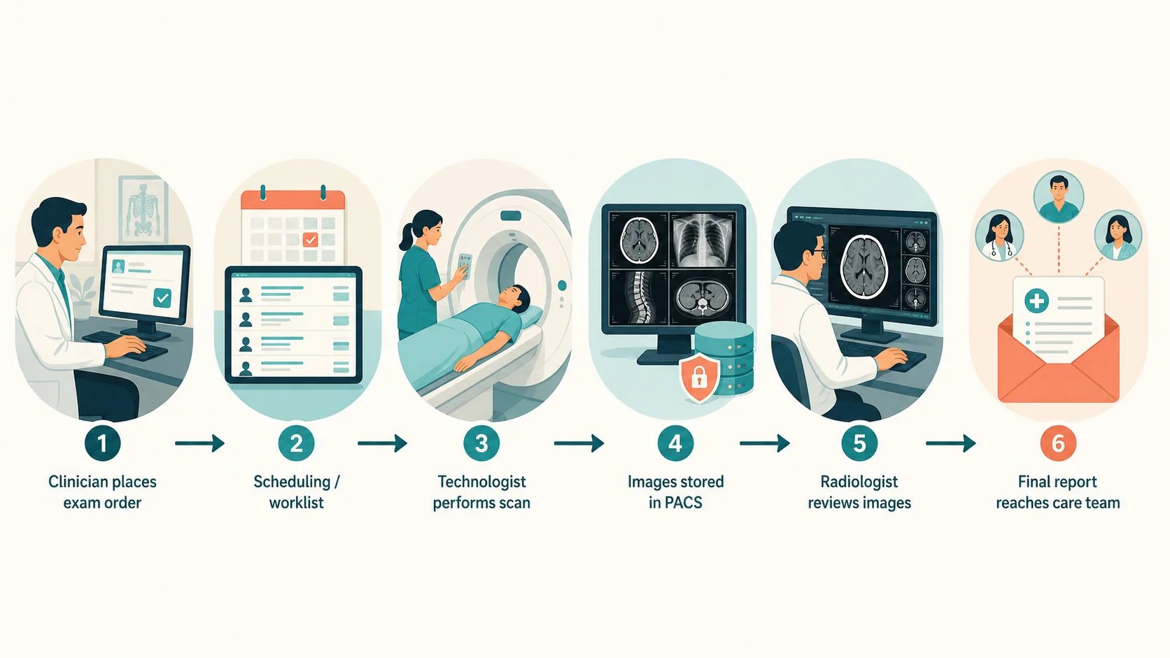
A visual guide to the path from an imaging order through acquisition, PACS, interpretation, and report delivery.

A two-page visual guide covering axial, coronal, and sagittal imaging planes alongside AP, PA, lateral, and oblique X-ray projections.

A side-by-side learning guide showing how matching patient and accession data keeps imaging moving, and where broken handoffs create delays and risk.

A visual guide to how imaging studies reach an AI server, how results can return to the clinical workflow, and why the output format must match the radiologist's needs and connected systems.
A combined guide covering the imaging project lifecycle and practical testing, readiness, and support considerations for go-live.