Basics of Imaging Informatics
How images, data, standards, systems, and people connect - PACS, viewers, DICOM, HL7, worklists, and reporting, explained at a high level.
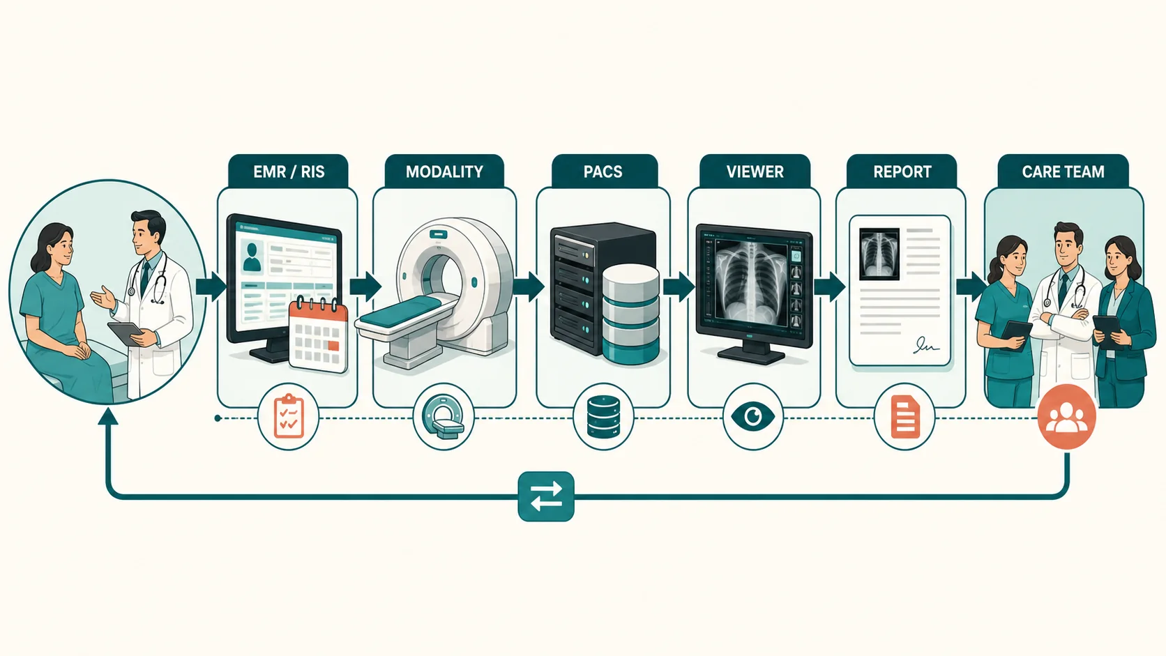
Imaging informatics is the field that connects medical imaging, information systems, clinical workflow, and technology. It sits between clinical imaging and healthcare IT, and its job is to make sure imaging information moves correctly, displays correctly, and supports patient care.
A working definition: imaging informatics helps the right images and the right information reach the right people at the right time.
Why the field exists
An imaging exam is far more than pixels. Around every study sits patient demographics, order and procedure details, modality information, study status, prior exams, measurements, reports, workflow events, and user activity - and that information travels between several systems.
When something is missing, late, mismatched, or sent to the wrong place, the workflow breaks in recognizable ways: a patient who does not appear on the modality worklist, images that never reach PACS, a study missing from the reading list, priors that will not load, or a report that does not return to the right system. Preventing, finding, and fixing those problems is the daily work.


Two standards beginners will meet
DICOM is used to store and exchange medical images. It carries both the image data and information about the patient, study, and exam. HL7 is used to exchange healthcare information such as patient demographics, orders, and results. Together, these standards help different healthcare systems communicate with each other.
The cast of systems and standards
- PACS (Picture Archiving and Communication System) - stores images and makes them available to view, compare, and interpret.
- Image viewer - the software clinicians actually look at studies in (zoom, pan, window/level, measurement, comparison). For most users it is the imaging system.
- RIS (Radiology Information System) - manages orders, scheduling, and the radiology record.
- Worklists - organized lists of work. A modality worklist puts the right patient and exam in front of the technologist; a reading worklist puts the right studies in front of the radiologist.
- Reporting system - where the final interpretation is created, then sent back to the care team.
Related free videos
DICOM Is Not Just Images (And It’s Not Software Either)
A practical overview of what DICOM really is in the imaging environment.
Why Do We Need the DICOM Header? (Explained in 3 Minutes)
A short explanation of how metadata gives the image clinical meaning.
DICOM UIDs Explained Simply – The Foundation of PACS Communication
A beginner-friendly explanation of the identifiers imaging systems rely on.
PACS Storage Explained: Archive vs Cache vs Workstation
A clear companion lesson on where images sit and how PACS balances access and storage.
DICOM vs DICOMweb: What's Actually Different?
A concise comparison of traditional image exchange and newer web-based approaches.
Full Radiology Workflow Explained: EMR → RIS → DICOM → PACS → Final Report
A workflow companion that shows how the major systems connect from order to result.
A simple workflow
Behind that one line, systems are constantly exchanging messages, images, status updates, and results. Imaging informatics is what keeps those systems working together reliably.
What the role actually focuses on
Most of the work reduces to practical, checkable questions: did the order arrive correctly, did the patient reach the modality worklist, did the right exam get selected, did the images send, did the study and its priors display correctly, did it route to the right reading list, did the report return to the right place, and did the workflow match how the clinic actually works? Answering those takes both technical and operational understanding.
Key takeaway
Imaging informatics connects systems, data, standards, workflow, and people. It spans PACS, viewers, DICOM, HL7, worklists, reporting, routing, and image exchange. The point is not simply to store images - it is to support safe, efficient, reliable imaging care.
Continue learning
This is the overview. Structured training goes deeper into PACS architecture, DICOM, HL7, modality worklists, routing, troubleshooting, reporting workflows, enterprise imaging, and real operational scenarios.