Medical Imaging Foundations: A Practical Introduction
What medical imaging is for, why exams are ordered, and how each image becomes part of a patient's clinical record.
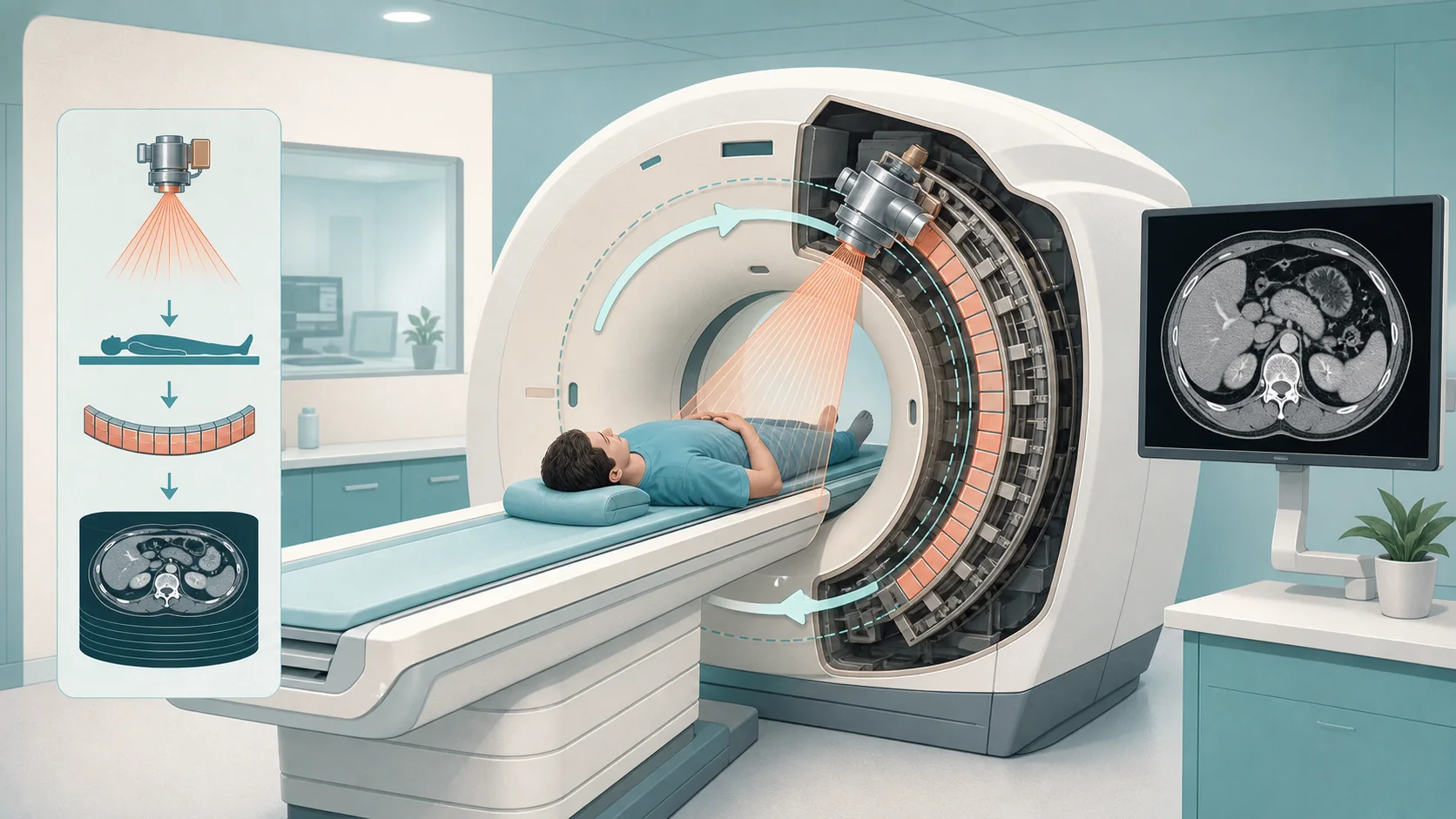
Medical imaging uses specialized equipment to create pictures of the inside of the body so that clinicians can see what they cannot examine directly. Those images help assess anatomy, find abnormalities, support a diagnosis, guide treatment, and track a condition over time.
At its simplest, imaging helps a care team answer three practical questions: what is wrong, where is it, and how serious is it?
An image is clinical information, not just a picture
This is the single most important idea for anyone entering imaging informatics. A medical image only has meaning when it stays attached to the information that describes it - the patient it belongs to, the exam that was performed, when and where, who ordered it, who interpreted it, and the final report.
A chest image with no patient, no date, and no context is not a diagnosis. It is an orphan. Keeping the image and its surrounding information correctly linked, from order to report, is most of what imaging informatics exists to do.
The common modalities, at a glance
Each imaging method ("modality") uses different physics and suits different questions:
Not all modalities work the same way. X-ray and CT use ionizing radiation; MRI and ultrasound do not. Different technologies are chosen because they answer different clinical questions, and some exams use a contrast agent to make certain anatomy or pathology easier to see.
- X-ray (radiography) - fast, widely available; bones, chest, abdomen.
- CT - X-rays plus computing to build detailed cross-sections of the body.
- MRI - magnetic fields and radio signals; excellent for soft tissue (brain, spine, joints).
- Ultrasound - sound waves, real-time, no ionizing radiation; abdomen, obstetrics, vascular.
- Mammography - dedicated breast imaging for screening and diagnosis.
- Nuclear medicine / PET - small amounts of radiotracer to show function, not just structure.
The right modality depends on the clinical question, the anatomy, the patient, and the kind of information needed. No single modality is "best."
The basic path: order to report
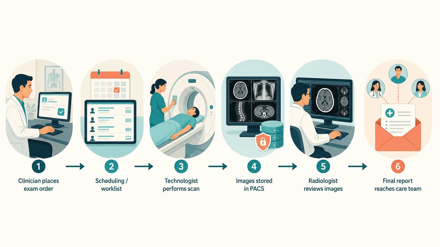
Most exams follow the same high-level path. A provider orders the exam; it is scheduled and appears as a task to image; a technologist performs it; the images are stored and made available; an interpreting physician reviews them and writes a report; the report goes back to the care team.
Several systems usually touch that path - the electronic record, scheduling, the modality, PACS, the reporting system, and the tools that distribute results.
Related free videos
Full Radiology Workflow Explained: EMR → RIS → DICOM → PACS → Final Report
A practical walk-through of the order-to-report path that supports this foundations article.
What Is WORKFLOW? | A Simple Explanation for Medical Imaging
A short companion lesson on how workflow connects the patient, exam, images, and report.
Why this matters for informatics
Before PACS, DICOM, HL7, and worklists make sense, it helps to hold the simple picture in mind: the patient, the order, the exam, the images, the interpretation, and the report all have to stay connected as they move between systems. Imaging informatics is the discipline that keeps that true.
Key takeaway
Medical imaging produces clinical information in the form of images plus the data that gives them meaning. Those pieces must stay linked to the correct patient, order, exam, and report. Imaging informatics supports the systems and workflows that make that happen.
Continue learning
This is the high-level map. Structured training goes deeper into how exams are ordered, performed, stored, displayed, interpreted, reported, and exchanged between systems.